
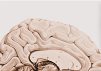

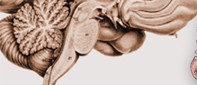


Anatomy
of the Pituitary Gland: The
pituitary gland is a small piece of tissue of dual origin (pharynx
for the adenohypophysis and brain for the neurohypophysis) attached
to the brain by a stalk. The pituitary gland hangs downward from
the brain and dwells in a small cavity known as the sella. The shape
of the cavity containing the pituitary gland mimics a Turkish horse
saddle (Sella Tursica). The gland is located at the center of the
head at the juncture between the cranial cavity and nasal cavity.
Embryonically, it is formed partly from the brain tissue itself
(posterior lobe) and partly the throat tissue (anterior lobe). Once
they are joined together during the developmental stage in a fetus,
it forms the pituitary gland as a whole. The cherry-shaped pituitary
gland is attached by a stalk (pituitary stalk) to the base of the
brain, the hypothalamus. The posterior lobe, also known as the neurohypophysis,
is directly connected to the hypothalamus by the pituitary stalk. Just above the pituitary gland lie the continuous visual nerve cables such as the right and left optic nerves, the optic chiasm which are the conjoined crossing cables, and the optic tracts which are the visual nerve cables conducting visual impulses to the brain. Just above the visual system is the hypothalamus, which is the vital brain tissue controlling homeostasis (maintenance of equilibrium-like conditions such as temperature and other parts of the autonomic nervous system, appetite, etc.) and instincts of the human body. |
|||||||||
----------------------------------------------------------------------------------------- |
|||||||||
Pituitary
Tumors: Many types of tumors can occur in the pituitary
gland. The most common tumors in the pituitary gland are pituitary
adenomas (which are benign). However, metastatic cancer can dwell
in the pituitary gland when the cancer cells metastasize to the
pituitary gland. Pituitary adenomas that produce ACTH cause Cushing's Disease. Symptoms of Cushing's disease include ruddy moon face, truncal obesity, buffalo hump, hypertension, abdominal striae, easy bruisability, depression, psychosis, irregular menses, impotence, osteoporosis, muscle weakness, etc. Cushing's disease is a serious condition requiring prompt treatments. Pituitary adenomas that produce growth hormone cause gigantism in children (due to still active growth plates) or Acromegaly in adults. In acromegaly the jaw, cheeks, fingers, and toes are thickened along with the enlargement of soft tissues such as the tongue, nose, and lips. Affected adult patients might find that their shoes or hats do not fit properly any longer. TSH-producing adenomas, which are relatively rare, cause goiters (enlarged thyroid gland) and thyroid hyper-function disorders. |
|||||||||
----------------------------------------------------------------------------------------- |
|||||||||
Symptoms
of Pituitary Tumors
Symptoms of pituitary tumors are: 1) , 2) , 3) , 4)
When functioning pituitary adenomas produce hormones, the excess amount of hormone will cause particular hormonal disorder symptoms depending on the type of hormone as described earlier. When pituitary adenomas become large, they compress and make the normal pituitary gland tissue hypo- or non-functional (hypopituitarism) resulting in pituitary hormonal dysfunction. Most often, nonfunctioning adenomas grow to large sizes undetected and result in hypopituitarism. However, functioning adenomas can also cause general hypopituitarism even while producing an excess amount of a particular hormone. When pituitary tumors compress the optic system (which is located just above the pituitary gland), visual disorders may develop. The most common visual disorder is a visual field defect at the outside view of each eye known as bitemporal hemianopsia. Other
symptoms such as memory disorder, hydrocephalus, and other
brain dysfunction can develop if the tumor is large enough to compress
the hypothalamus. Pituitary adenomas are known to bleed spontaneously
(pituitary apoplexy). When spontaneous bleeding occurs inside a
pituitary adenoma, symptoms of severe headaches, visual disorders,
eye movement disorder, and altered consciousness can develop. Immediate
medical treatment is necessary and surgical treatment is often required. |
|||||||||
----------------------------------------------------------------------------------------- |
|||||||||
|
Treatments of Pituitary
Tumors
Medical,
surgical, and/or radiation treatments are available for pituitary
tumors. Prolactinomas can be treated with medications
(bromocriptine is most commonly used). Surgical treatment is indicated
when medication side effects or intolerance develops. Acromegaly
can be treated with octreotide medication but does not respond as
well as prolactinomas do with bromocriptine. Often, acromegaly requires
surgical removal of the tumor. If surgery is not successful, octreotide
medication or radiation treatment (gamma-knife surgery) can be considered.
Cushing's disease requires surgical removal of
the tumor as the first line of treatment. If surgical treatments
fails to relieve symptoms, radiosurgery or medical treatments can
be instituted. TSH-secreting adenomas require surgical
treatment. When nonfunctioning tumors cause symptoms,
surgical treatment is the first choice among the treatments. If
surgical treatments are not successful, radiation treatment (conventional
radiation or radiosurgery) has to be considered. Endoscopic
Pituitary Surgery is performed through a natural nasal
air pathway without any incisions (unlike the conventional microscopic
surgery performed with an incision made under the upper lip or inner
aspect of a nostril). Endoscopic surgery does not require the use
of a metallic transsphenoidal retractor that is used for conventional
microscopic surgery. A 4-mm endoscope is placed in front of the
tumor in the sphenoidal sinus and the tumor is removed with specially
designed surgical tools. Conventional transsphenoidal surgery is
performed under the operating microscope via an incision underneath
the upper lip (sublabial incision) or intranasal incision (transfixional
incision). It requires two to three days of nasal packing and three
to five days of hospital stay. |
|||||||||